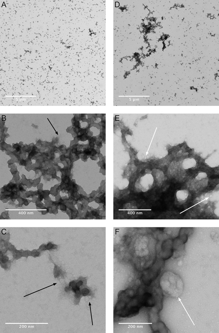
Figure 3.

Transmission electron micrographs show aggregate morphology after incubation of RPMI 1640 + 10% FBS with Ti (A‐C) or Ti + Co (D‐F) without the presence of macrophages. Magnification increasing from A to C and from D to F. Nano‐filament like structures are seen at the edges of the aggregates (C). Protein aggregates are larger and more electron dense with Ti + Co (D‐F) than with only Ti (A–C). Filament (B, C) and exosome (E, F) like structures indicated with black‐ and white arrows.
