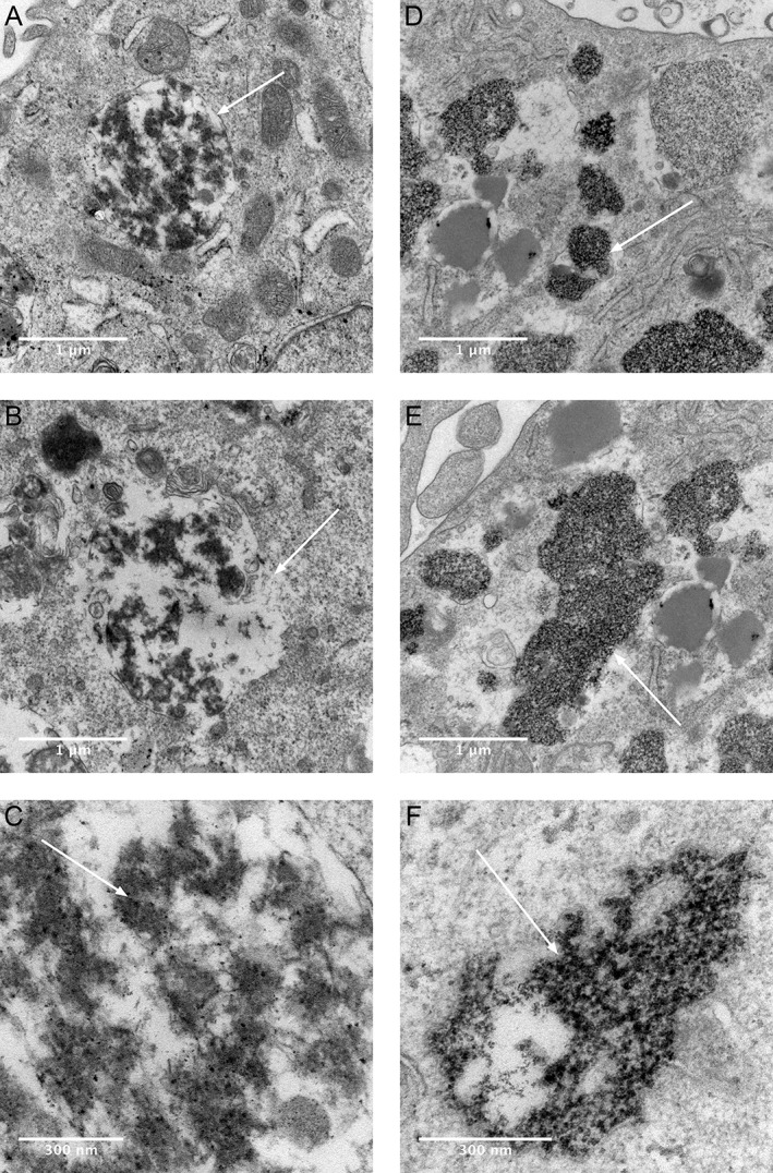
Figure 5.

Electron micrographs show aggregate uptake into human macrophage THP‐1 cells in vitro after exposure to Ti (A‐C) or Ti + Co (D‐F) with RPMI 1640 + 10% FBS medium. Transmission electron micrograph images are shown in two different magnifications. Uptake of Ti‐protein aggregates with a surrounding membrane (A, white arrow), and that structure shows some similarity to a phagolysosome. Destruction of the lysosomal membrane, which contains high‐density Ti aggregates (B, white arrow). Visible Ti nanoparticles inside lysosome (C, white arrow). Ti‐Co protein aggregates are more concentrated, located freely in the cytoplasm, and have no visible surrounding membrane (D–F) compared with Ti aggregates (A–C). High density cluster of metal aggregates inside the cytoplasm (D, white arrow). No visible membrane surrounding the metal‐rich cluster of protein aggregates (E, F, white arrow).
