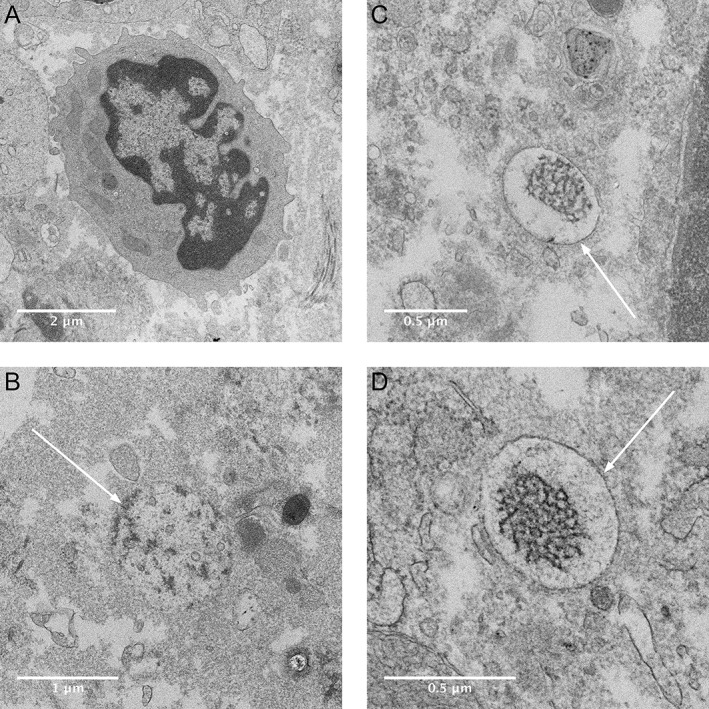
Figure 6.

Electron micrographs showing phagocyte and intracellular aggregates in human peri‐implantitis biopsy. Host cell with macrophage like structures (A). Intracellular structures with electron‐dense content with various membrane structure (indicated with arrows) (B–D) similar to that found in the THP‐1 cells exposed to Ti RPMI 1640 + 10% FBS. No similar structures were found in the sections of tissue from the periodontitis biopsy (not shown).
