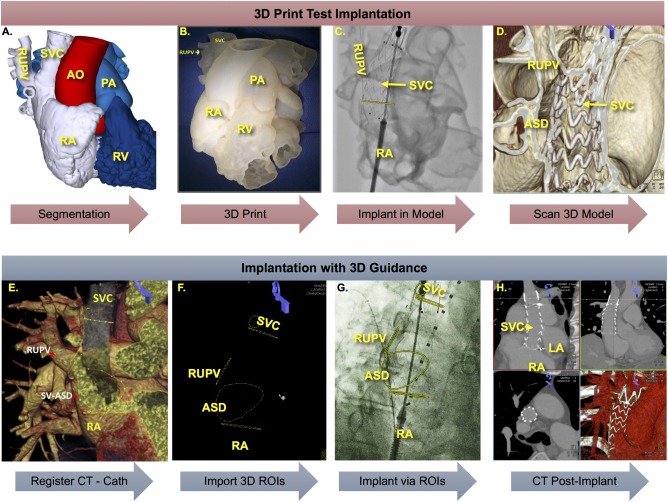
Figure 3.

Simulation of device implantation in 3D printed model and implantation in patient using 3D image guidance. 3D segmentation of gated cardiac CTA in MIMICS (A) followed by 3D print of segmented structures of interest (B). Implantation of candidate stent graft in 3D printed model under fluoroscopy (C) and cone‐beam CT of results (D) demonstrated successful exclusion of ASD and RUPV. 3D guidance of implant in patient after importing gated cardiac CTA and registration with cone‐beam CT of patient on table (E) followed by overlay of ROIs onto fluoroscopy (F). G, Implant of stent graft guided by 3D ROIs demarcating the ASD and RUPV with proximal and distal ends of SVC outlined in orthogonal planes. H, Post‐procedure gated CTA demonstrated successful exclusion of ASD and RUPV without obstruction. [Color figure can be viewed at http://wileyonlinelibrary.com]
