Figure 5.

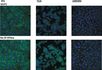
Immunofluorescence confocal images (63X). Cells were stained for GLUT‐1 (upper panels) and Na+/K+ ATPase (bottom). Nuclei were counterstained with DAPI (blue).

Immunofluorescence confocal images (63X). Cells were stained for GLUT‐1 (upper panels) and Na+/K+ ATPase (bottom). Nuclei were counterstained with DAPI (blue).