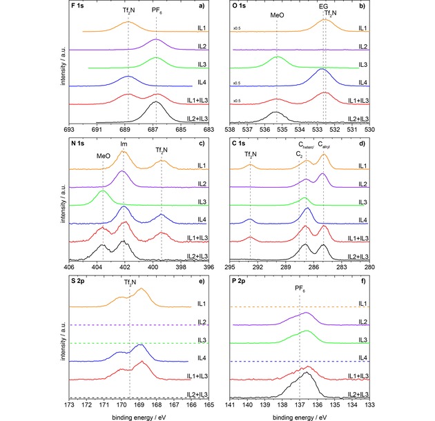
Figure 1.

F 1s, O 1s, N 1s, C 1s, S 2p, and P 2p spectra of the investigated ILs and IL mixtures in 0° emission, measured with Al Kα radiation. Orange: IL1, [C8C1Im][Tf2N]; violet: IL2, [C8C1Im][PF6]; green: IL3, [(MeO)2Im][PF6]; blue: IL4, [Me(EG)2C1Im][Tf2N]; red: equimolar mixture of IL1+IL3, [C8C1Im][Tf2N]+ [(MeO)2Im][PF6]; black: equimolar mixture of IL2+IL3, [C8C1Im][PF6]+[(MeO)2Im][PF6]. All spectra are normalized to the fitted peak intensity of the F 1s spectrum of [(MeO)2Im][PF6]. Abbreviations: MeO for [(MeO)2Im]+, Im for N 1s of [C8C1Im]+ and [Me(EG)2C1Im]+, EG for O 1s of [Me(EG)2C1Im]+, PF6 for [PF6]−, Tf2N for [Tf2N]−.
