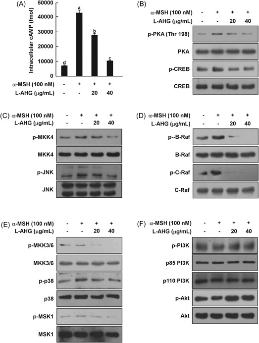
Figure 5.

Effect of l‐AHG on the α‐MSH‐induced melanogenic signaling pathway in HEMs. l‐AHG inhibited α‐MSH‐induced (A) cAMP levels (B) PKA/CREB, (C) JNK (D) ERK, and (E) p38 signaling in HEMs. Cells were treated with l‐AHG (20 or 40 μg/mL) for 1 hour before being exposed to α‐MSH (100 nM) and harvested 15 minutes later. (F) l‐AHG activated the α‐MSH‐induced dephosphorylation of Akt signaling. Cells were treated with l‐AHG (20 or 40 μg/mL) for 1 hour before being exposed to α‐MSH (100 nM) and harvested 30 minutes later. cAMP data (n = 3) represent the mean values ± SD. Means with letters (a‐d) in a graph are significantly different from each other at P < .05. Western blot analysis was performed using specific antibodies against the respective phosphorylated and total proteins. The data are representative of 3 independent experiments that yielded similar results. α‐MSH, alpha‐melanocyte‐stimulating hormone; cAMP, cyclic adenosine monophosphate; CREB, cAMP response element‐binding protein; ERK, extracellular signal‐regulated kinase; HEMs, human epidermal melanocytes; JNK, c‐Jun N‐terminal kinase; l‐AHG, 3,6‐anhydro‐l‐galactose; PKA, protein kinase A; SD, standard deviation
