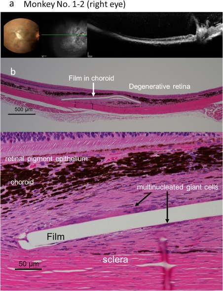
Figure 5.

Fundus photographs and optical coherence tomography (a), and pathology (b) of the right eye of Monkey No. 1–2 with 1‐month implantation of a dye‐coupled film. Horizontal section (green line) of optical coherence tomography is shown in the middle black‐and‐white fundus photograph. The film is not detected by optical coherence tomography and is later located in the choroid at pathology. Hematoxylin‐eosin stain. Scale bar = 500 µm (upper panel of b) and 50 µm (lower panel of b).
