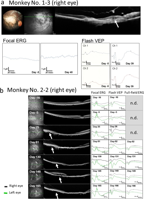
Figure 6.

(a) Focal electroretinograms (ERG) and visual evoked potential (flash VEP) in the right eye of Monkey No. 1–3 with 1‐month dye‐coupled film implantation. Focal ERG is extinguished after the induction of macular degeneration (Day −5) and also not detected at 1 month of film (white arrow in optical coherence tomography in a) implantation (Day 40). In contrast, reduced VEP amplitude at Day −5 becomes larger at 1 month of film implantation (Day 39). Channel 1 (Ch 1) and Channel 2 (Ch 2) correspond to the right and left visual cortex recordings, respectively, in photic stimuli to the right eye. (b) Focal ERG, flash VEP, and full‐field ERG in the right eye of Monkey No. 2–2 with 6‐month dye‐coupled film (white arrows in optical coherence tomography) implantation. Focal ERG is noted before the induction of macular degeneration (Day −16), extinguished after the development of macular degeneration (Day −5) and also not detected throughout the 6‐month course until Day 165. In contrast, reduced VEP amplitude on Day −5 becomes larger at 1 month and 6 months of film implantation (Day 26 and Day 166). Full‐field ERG which was scheduled to measure only from Day 82 shows normal waves throughout the course (n.d., not determined). Right eye recordings (black lines) of VEP are from the right visual cortex (Channel 1) in response to right eye photic stimuli while left eye recordings (green lines) are from the left visual cortex (Channel 2) in response to left eye stimuli. Vertical line (amplitude) and horizontal line (time) are 1 μV and 20 msec in focal ERG, 10 μV and 50 msec in flash VEP, and 100 μV and 50 msec in full‐field ERG, respectively. Note that space between film and retinal pigment epithelium appears temporarily on Day 148 in optical coherence tomography.
