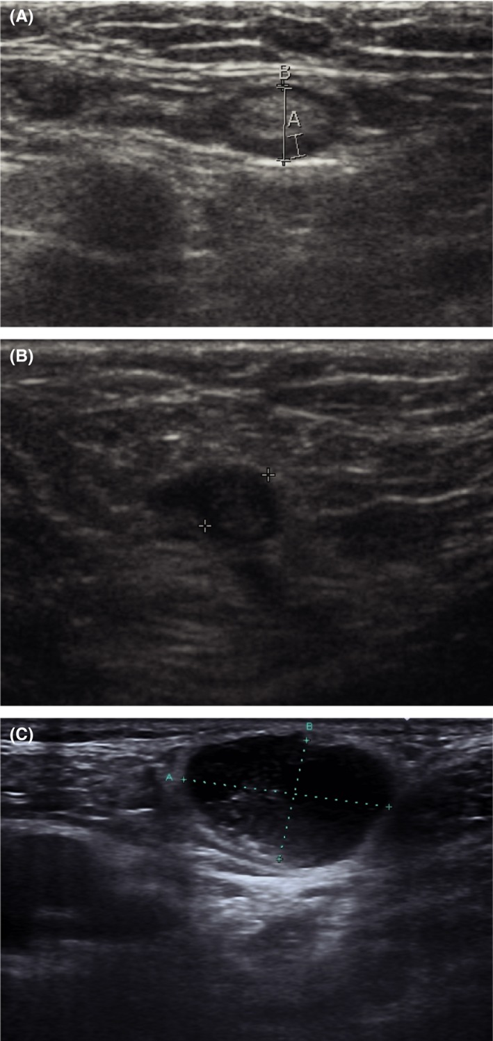
Figure 3.

Ultrasound images. (A) Ultrasound image of a normal lymph node in the groin (measurement A: 1.4 mm, B: 4.6 mm). (B) Ultrasound image of a metastatic lymph node (patient A): enlarged (diameter 5.9 mm), focal cortical thickening on the left side and loss of echogenic hilar sinus fat. (C) Ultrasound image of a metastatic lymph node (patient B): enlarged oval‐shaped, loss of echogenic hilar sinus fat (measurement A: 23.1 mm, B: 14.3 mm).
