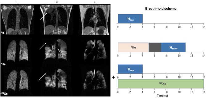
Figure 1.

Left: Comparison of 3He and 129Xe MR ventilation images of i. a healthy nonsmoker (group b), ii. a patient with NSCLC (white arrows indicate the location of a lesion), and iii. a patient with COPD. (Note: The slice thickness of 3He images is half that of 129Xe images; see Table 2). Right: Corresponding breath‐hold scheme for ventilation imaging scans. Scans were acquired in the order shown, with a change of RF coil and repositioning of the patient between 3He and 129Xe scans.
