Figure 2.

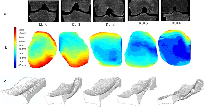
(a) Middle coronal slices of the VOI for different knee specimens with various KL classifications are represented with their corresponding maps. (b) 3D map of medial compartment with color scale from 0 to 5 mm. (c) The 3D joint space mask.

(a) Middle coronal slices of the VOI for different knee specimens with various KL classifications are represented with their corresponding maps. (b) 3D map of medial compartment with color scale from 0 to 5 mm. (c) The 3D joint space mask.