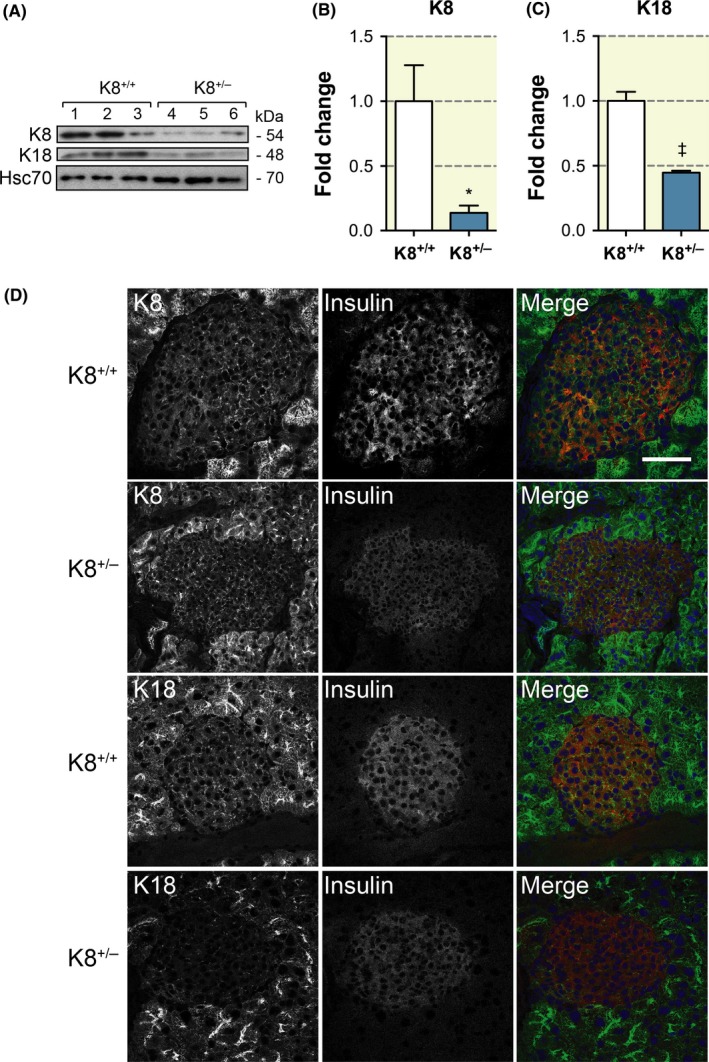
Figure 1.

K8+/− mouse islet cells express less K8 and K18 than K8+/+ islets. A, Western blot for K8, K18 and Hsc70 (loading control) of protein lysate from pancreatic islets isolated from K8+/+ and K8+/− mice (lane 1‐3, K8+/+; lane 4‐6, K8+/−). B, Quantification of immunoblotting showed a statistically significant fivefold decrease in K8 protein levels in K8+/−‐isolated pancreatic islets compared to K8+/+ islets when normalized to Hsc70 levels. *P < .05, error bars represent mean ± SEM. C, Quantification of immunoblotting showed a significant twofold decrease in K18 protein levels in K8+/−‐isolated pancreatic islets compared to K8+/+ islets when normalized to Hsc70 levels. †P < .001, error bars represent mean ± SEM. D, Immunostaining of K8 or K18 (green), insulin (red, used as a marker for islets) and nuclei (blue, colours seen in the merged images) of pancreatic sections from K8+/+ and K8+/− mice shows a less dense islet keratin network in K8+/−.mice compared to K8+/+ mice. Scale bar: 100 μm
