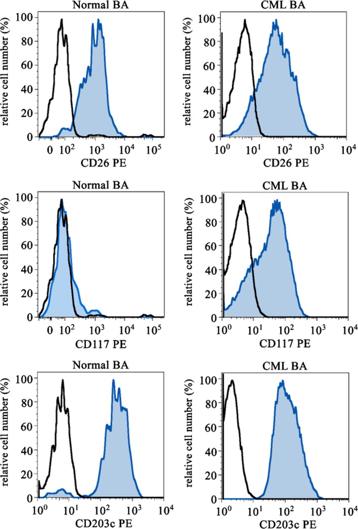
Figure 1.

Expression of cell surface antigen on blood basophils. Peripheral blood basophils of healthy normal donors (left images) and of patients with chronic myeloid leukaemia (CML, right images) were stained with antibodies against CD26 (upper panels), CD117 (KIT) (middle panels) and CD203c (lower panels) by multicolour flow cytometry. Basophils were detected by their typical side scatter characteristics, expression of CD123 and CD203c and exclusion of CD34 positivity
