Figure 1.

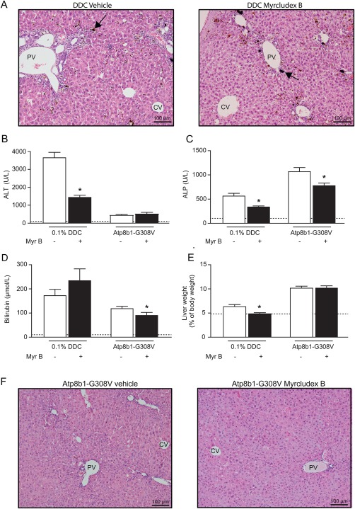
Reduced cholestatic liver injury by myrcludex B in DDC and PFIC1 models. (A) Liver H&E staining showing vehicle and myrcludex B treatment in DDC‐fed wild‐type mice. Black arrows indicate porphyrin plugs. (B‐C). Plasma biochemistry displaying degree of parenchymal liver injury (ALT in U/L) and cholestasis (ALP in U/L) in DDC‐fed mice and Atp8b1‐G308V mice. Dotted lines indicate physiological levels of the indicated enzymes. (D) Total bilirubin levels in plasma in DDC‐fed mice and Atp8b1‐G308V mutant mice. (E) Liver to BW ratio in DDC‐fed mice and Atp8b1‐G308V mice. (F) Liver H&E staining of Atp8b1‐G308V mice treated with vehicle or myrcludex B, compaction of hepatocytes is noted around periportal areas. Asterisk indicates significant changes (n=8‐14 mice/group). Abbreviations: CV, central vein; PV, portal vein.
