Figure 1.

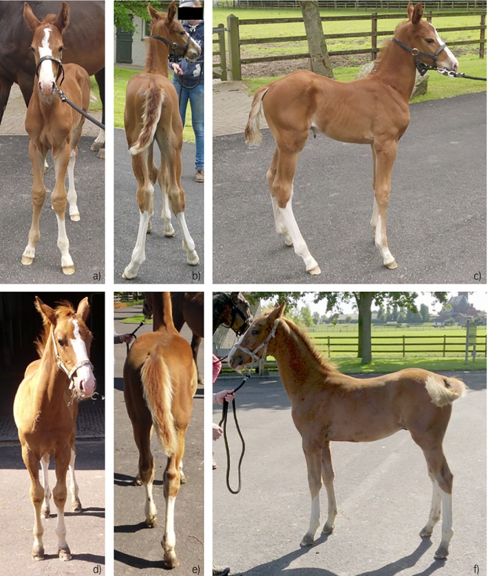
Front a) & d), hind b) & e) and lateral c) & f) views of the same foal at the age of 1 week a), b) and c) and 12 weeks d), e) and f) illustrating conformational changes over time. Note the subtle valgus deviation at week 1 in all 4 limbs.

Front a) & d), hind b) & e) and lateral c) & f) views of the same foal at the age of 1 week a), b) and c) and 12 weeks d), e) and f) illustrating conformational changes over time. Note the subtle valgus deviation at week 1 in all 4 limbs.