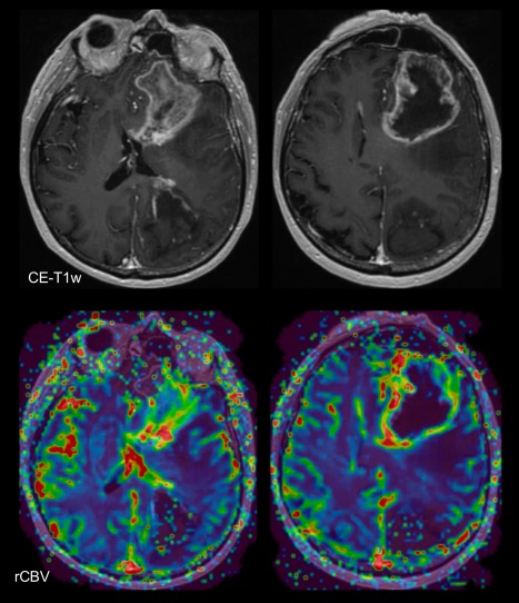
Figure 5.

MRI features of histopathologically proven glioblastoma: Contrast‐enhanced (CE) T1‐weighted (T1w) sequences shows an enhancing lesion in the left frontal lobe, with areas of central necrosis. There is increased rCBV (green/red) in the enhancing tumor portions. Note there also hemorrhage in the left parieto‐occipital region.
