Figure 6.

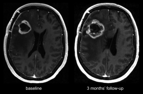
Histopathologically confirmed pseudoprogression, where postcontrast T1‐weighted images show a “swiss cheese” or “soap bubble” increase of the margin of the lesion in the right frontal lobe.

Histopathologically confirmed pseudoprogression, where postcontrast T1‐weighted images show a “swiss cheese” or “soap bubble” increase of the margin of the lesion in the right frontal lobe.