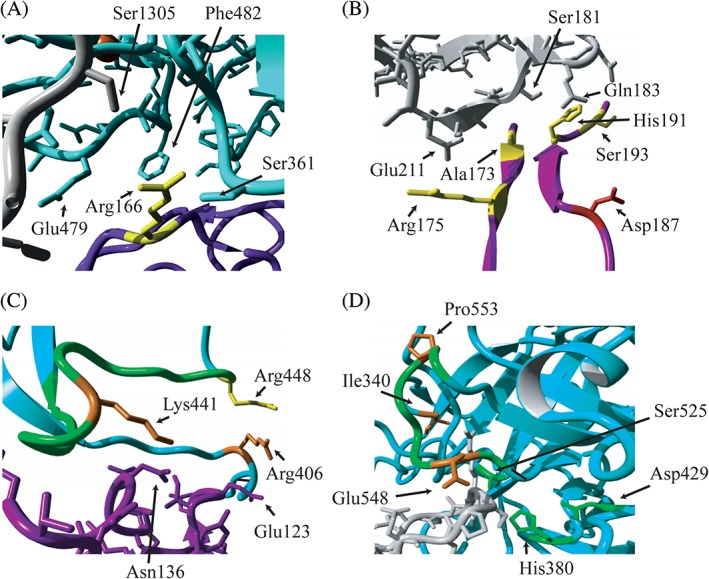
Figure 3.

Localization of altered residues on the interface of C3b, FH and FI in proteins domains containing a higher percentage of alleles in aHUS/C3G or AMD. Fragments of the 3‐dimensional structure of C3b (gray), FI (cyan) and FH construct (purple) are shown. The residues altered in the SCR3 domain of FH (A and B) and the SP domain of FI (C and D) are shown. Residues that were found mutated in AMD only (yellow), aHUS/C3G (red) or both phenotypes (orange) are indicated, as well as amino acids of interacting partners that are in close proximity of the mutated residues (A, B, C). Important structural elements of FI are indicated in green: the charged loop 435‐448 (C); the activation loop 548‐553 and the catalytic triad (H380, D429, S525) (D). The figure is generated based on the PDB 5O32,29 using YASARA Version 17.8.1531
