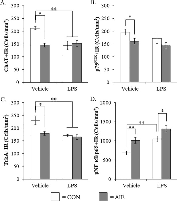
Fig 7. Lipopolysaccharide (LPS) treatment mimicked the adolescent intermittent ethanol (AIE)-induced loss of cholinergic neuron markers and increased phosphorylation of the proinflammatory transcription factor nuclear factor kappa-light-chain-enhancer of activated B cells p65 (phosphorylated [pNF-κB p65]) in the adult male basal forebrain.
(A) Modified unbiased stereological assessment of choline acetyltransferase (ChAT)+IR neurons in the basal forebrain of adult (P80) rats revealed a significant reduction in AIE- (31% [±4]), CON+LPS- (32% [±8%]), and AIE+LPS-treated animals (28% [±5%]), relative to CONs. (B) Modified unbiased stereological assessment of p75NTR+IR neurons in the basal forebrain of adult (P80) rats revealed a significant reduction in AIE-treated animals (18% [±5]) while LPS treatment did not alter expression of p75NTR. (C) Modified unbiased stereological assessment of tropomyosin receptor kinase A (TrkA)+IR neurons in the basal forebrain of adult (P80) rats revealed a significant reduction in AIE- (22% [±5]), CON+LPS- (26% [±2]), and AIE+LPS-treated animals (28% [±5]), relative to CONs. (D) Modified unbiased stereological assessment of pNF-κB p65+IR cells in the basal forebrain of adult (P80) rats revealed a significant increase in AIE- (48% [±11%]), CON+LPS- (54% [±11%]), and AIE+LPS-treated animals (93% [±12%]), relative to CONs. Data are presented as mean ± SEM. * p < 0.05, ** p < 0.01.

