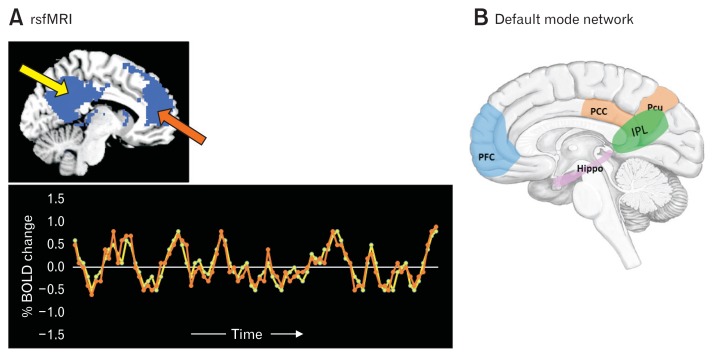
Figure 3.
Resting-state functional magnetic resonance imaging (rsfMRI) and default mode network. (A) rsfMRI is used to investigate synchronous neural activity (as measured with the blood oxygen level-dependent [BOLD] signal) between spatially distinct brain regions and provides the functional architecture of the brain. The lower panel represents the synchronous fMRI BOLD signal activity from the posterior cingulate cortex (yellow arrow in the upper panel) and in the medial prefrontal cortex (orange arrow in the upper panel). Adapted from Raichle.58 (B) Default mode network (DMN): the set of areas that work together at rest and are involved in high-level cognitive processes such as self-awareness and memory. DMN is thought to consist of the medial prefrontal cortex (mPFC), posterior cingulate cortex (PCC), hippocampus (Hippo), superior temporal gyrus, inferior parietal lobule (IPL), and precuneus (Pcu).

