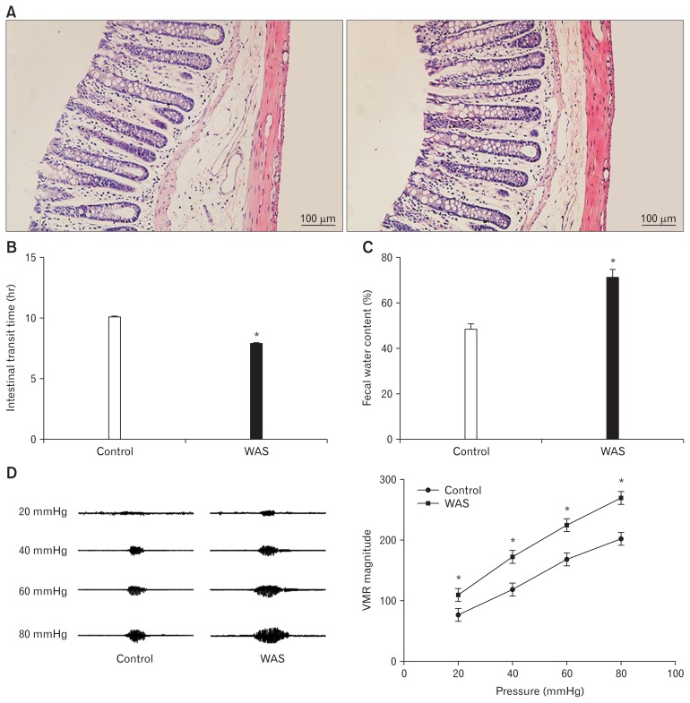
Figure 1.
Evaluation of the animal model. (A) Hematoxylin-eosin staining (×200) showing that colon specimens of the control (left) and WAS (right) groups are intact without congestion and obvious infiltration of inflammatory cells. (B) The intestinal transit time of water avoidance stress (WAS) rats is shorter than that of control rats. (C) The fecal water content in the WAS group is increased compared to that in the control group. (D) Visceromotor response (VMR) cure (left) and summary data (right) for VMR responses to CRD at pressures of 20, 40, 60, and 80 mmHg in control and WAS rats. The VMR amplitude of WAS rats is significantly higher than that of control rats. *P < 0.05 compared to controls.

