. 2018 Sep 26;7:e37881. doi: 10.7554/eLife.37881

Figure 6. Par3 recruits Pk3 to the apical side of the cell in vivo.

Embryos were injected with GFP-Pk3 and Myc-Par3 RNAs (100 pg each), cryosectioned at stage 10.5 and immunostained with indicated antibodies (A) Scheme showing a relative position of imaged superficial ectoderm cells. Both endogenous Par3 (B), and exogenous Myc-Par3 (C, E’, E’’) are apically localized. (C’) Lack of Myc-Par3 staining with anti-GFP antibody. (D) Lack of apical enrichment of exogenous GFP-Pk3. (E–E’’) Myc-Par3 recruits GFP-Pk3 to the apical surface. (B, C, E) Apical enrichment is shown by arrowheads. The apical recruitment of Pk3 was observed in > 90% of cells coinjected with Pk3 and Par3. Each group contained five embryos. The same results were obtained in five independent experiments. Scale bar 10 µm.

Figure 6.

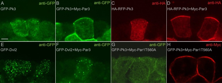
Figure 6—figure supplement 1. Par3 recruits Pk3 but not Dvl2 to the apical cell membrane.

Figure 6—figure supplement 1.

8–16 cell Xenopus embryos were injected animally with 100 pg of the RNAs encoding GFP-Pk3 (Pk3), HA-RFP-Pk3, GFP-Dvl2, Myc-Par3 and Myc-Par1T560A as indicated, cultured until stage 10.5, cryosectioned and immunostained for GFP (A, B, E–G), HA (C, D) or Myc (H). Myc-Par3 recruits GFP-Pk3 (A, B), but not GFP-Dvl2 (E, F), to the apical membrane. (C, D) Myc-Par3 promotes the apical localization of HA-RFP-Pk3. (G) Myc-Par1T560A does not promote GFP-Pk3 apical localization. (H) Myc-Par1T560A is cortically localized. Scale bar, 10 µm. The apical recruitment of Pk3 was observed in the majority (>90%, n > 100) of cells co-expressing Pk3 and Par3, but not in cells expressing Pk3 alone.