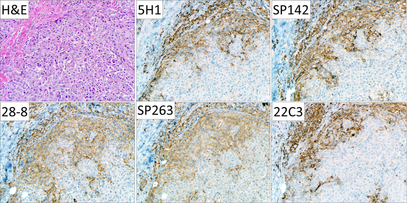
Figure 1. Representative photomicrographs of PD-L1 immunohistochemistry performed on a cutaneous melanoma metastasis using clones 5H1, SP142, 28-8, SP263, and 22C3.
All five antibodies showed similar regional patterns of PD-L1 expression by tumor cells, macrophages, and lymphocytes. A degree of geographic heterogeneity between the different tissue sections is evident. Qualitative differences in staining with regard to cytoplasmic or membranous staining or non-specific background staining were not observed. Original magnification, 200x, all fields.

