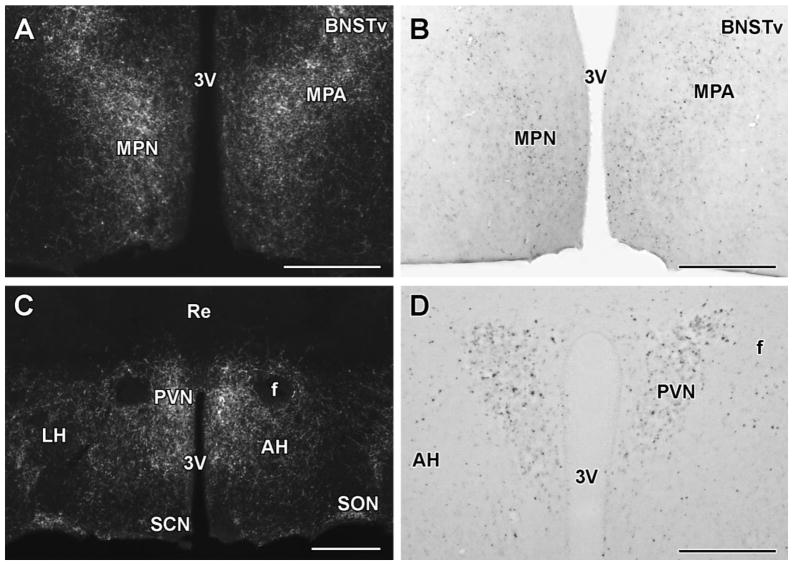
Fig. 3.
TIP39-ir fibers and parathyroid hormone 2 receptor (PTH2 receptor)-expressing neurons in the preoptic and anterior hypothalamus of mice. A: TIP39-ir fibers are abundant in the medial preoptic nucleus (MPN) as well as dorsolateral to it in the medial preoptic area (MPA) and ventral subdivision of the bed nucleus of the stria terminalis (BNSTv). B: X-gal histochemistry in a mouse expressing LacZ driven by the promoter of the PTH2 receptor. The PTH2 receptor-expressing neurons are distributed similarly to TIP39 fibers. C: TIP39-ir fibers have particularly high density in the paraventricular hypothalamic nucleus (PVN). They are also present in the anterior and lateral hypothalamic areas (AH and LH, respectively) albeit with only a low density in the LH. TIP39 fibers are present in the supraoptic (SON) but absent in the suprachiasmatic nucleus (SCN). D: PTH2 receptor-expressing neurons are abundant in the PVN. Scale bar = 400 μm for A and B, 500 μm for C, and 300 μm for D. The figure is created from the material presented previously (Faber et al., 2007).

