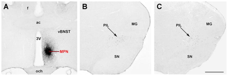
Fig. 4.
Comparison of neurons in the PIL retrogradely labeled from the medial preoptic nucleus and those that express c-Fos in response to pup exposure in mothers. A: Injection site of the retrograde tracer, cholera toxin beta subunit, in the medial preoptic nucleus (MPN). B: Retrogradely labeled neurons in the posterior intralaminar complex of the thalamus (PIL). C: Fos-immunoreactive neurons in the PIL following suckling in mother rats. Additional abbreviations: ac–anterior commissure, f–fornix, och–optic chiasm, MG–medial geniculate body, SN–substantia nigra, vBNST – ventral subdivision of the bed nucleus of the stria terminalis. Scale bar = 1 mm. The figure is created from the material of our previous paper (Cservenak et al., 2017b).

