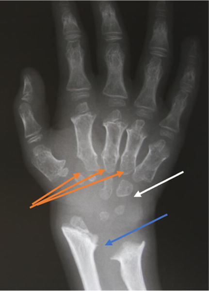
Figure 9.

X-ray radiograph of an MPS IVA patient hand. Clearly seen are the tapering of the proximal portion for the metacarpals (orange arrows), small irregular carpal bones (white arrow), and the distal portion of the radius being tilted toward the ulna (blue arrow).
