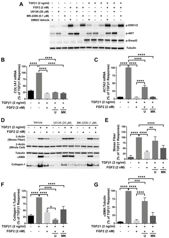
Figure 6.
FGF2 inhibition of TGFβ1 induced αSMA requires MEK/ERK, but not AKT. Primary HLFs were serum-starved and treated with FGF2 (2 nM), TGFβ1 (2 ng/ml), or FGF2 + TGFβ1 for 1 hour or 48 hours in the presence or absence of U0126 (20 μM) or MK-2206 (0.1 μM). (A) Total protein was collected at 1 hour and immunoblotting was performed for phosphorylated ERK1/2, AKT, and Smad2, and normalized to tubulin. (B, C) Total RNA was collected at 48 hours, and qRT-PCR was performed for COL1A1 (B) or ACTA2 (C). ΔCt values were normalized to GAPDH and expressed as % of TGFβ1 response. (D) Purified stress fibers and total protein were collected at 48 hours, and immunoblotting was performed for beta-actin, αSMA, collagen-1, and tubulin. (E–G) Densitometry was performed and expressed as % of TGFβ1 response. * indicates p < 0.05, ** indicates p < 0.01, *** indicates p < 0.005, **** indicates p < 0.001 using an unpaired 2-way t-test.

