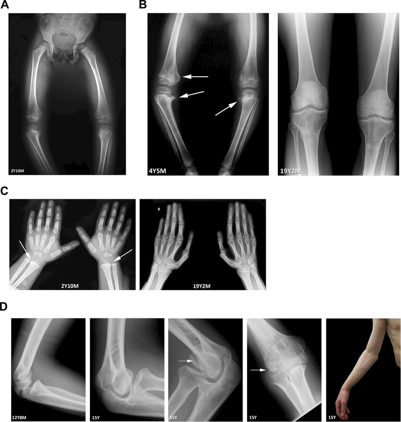
Figure 2: Radiographs of long bones and joints.
(A) Bilateral genu varum, first noted at approximately 1 year of age, was first imaged at age 2 years 10 months. (B) Bilateral medial corner fractures and beaking in the distal femur and proximal tibiae (left, arrows) were first noted at age 4 years 5 months. These changes are reversed after surgical intervention and upon skeletal maturity (right). (C) Bilateral radial corner fractures in the wrist, first imaged at age 2 years 10 months (left, arrows) are no longer present upon skeletal maturity at age 19 years 2 months (right). (D) Boney changes (left) and limited extension (far right) of the elbow are suggestive of avascular necrosis.

