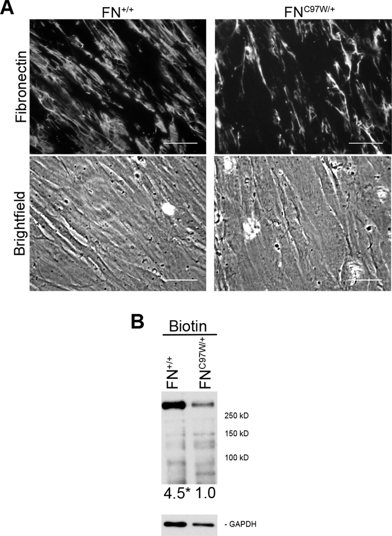
Figure 3: Primary FNC97W/+ cells assemble reduced FN matrix compared to primary FN+/+ cells.
(A) The FN matrix of wild type (FN+/+) and mutant (FNC97W/+) cells was visualized by immunofluorescence microscopy after staining with R184 anti-FN polyclonal antiserum (top). Equivalent cell densities are visible by phase microscopy (bottom). Total fluorescence averaged over 10 images was 191+34 for FNC97W/+ cells and 267+54 for FN+/+ cells (SD, p < 0.01). Scale bar = 40 μm. (B) Extracellular proteins of confluent cells were biotinylated using a cell-impermeable crosslinker, 125 ng of total protein was separated by SDS-PAGE, and blots were probed with HRP-coupled streptavidin (top). Relative levels of biotinylated FN are, on average, at least 4.5 for FNC97W/+ versus 1.0 for FN+/+ (p<0.01), after normalization to GAPDH as a loading control (bottom).

