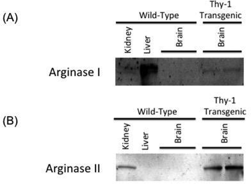
Figure 2.

(A) and (B) Western blots showing overexpression of arginase I or arginase II expression in brain lysates from Thy1-ArgI and Thy1-ArgII mice respectively as compared to WT mouse brain lysate. Liver and kidney tissues were used as positive controls for Arginase I and Arginase II respectively.
