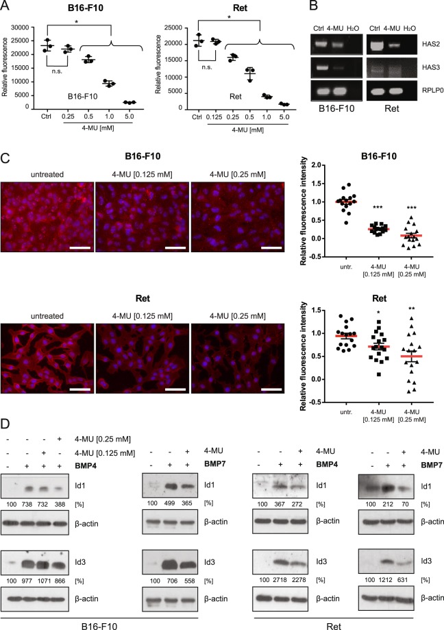
Figure 2.
Inhibition of HA synthesis reduces BMP-dependent Id1/3 protein expression in murine melanoma cells. (A) B16-F10 and Ret cells were treated with the indicated concentrations of 4-methylumbelliferone (4-MU) for 48 hours. The number of cells was then quantified on the basis of DNA content using CyQUANT assay. The mean and SE of triplicate samples are shown. Data represents means ± SD, n = 3. Student’s t-test: *p < 0.05; n.s., non-significant. (B) B16-F10 and Ret cells were treated with 0.25 mM (B16-F10) and 0.125 mM (Ret) 4-MU for 48 hours. Total RNA was prepared, and RT-PCR was performed to analyze the gene expression of HAS2 and HAS3. For a negative control, water instead of cDNA was added to the PCR reaction (H2O). Rplp0 was used as an internal loading control. (C) B16-F10 and Ret cells were treated or not treated with the indicated concentrations of 4-MU for 48 hours. Hyaluronic acid was detected using HABP immunofluorescence staining. HABP staining intensity was quantified using ImageJ. At least 15 images were quantified in a blinded fashion. Scale bar 50 μm. Student’s t-test: *p < 0.05, **p < 0.01, ***p < 0.001. (D) B16-F10 and Ret cells were treated or not treated with 4-MU (0.25 mM, B16-F10, right; 0.125 mM, Ret) for 48 hours, then stimulated with 10 ng/ml recombinant BMP4/7 for another 6 hours. Cell lysates were then analyzed for Id1/3 protein expression by Western blot. β-actin was used as a loading control. Densitometric quantification is shown below the Western blots, representing the signal normalized to the loading control (β-actin), relative to the untreated controls.

