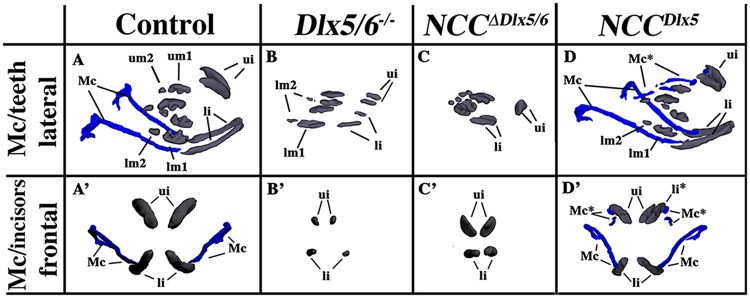
Figure 4.
Teeth complement and Meckelian cartilage from 3D reconstructions of control, Dlx5/6−/−, NCC∆Dlx5/6 and NCCDlx5 E18.5 mouse foetuses. Lateral (A–D) and frontal (A’–D’) views of teeth (grey) and Mekelian cartilage (blue) from control (A,A’), Dlx5/6−/− (B,B’), NCC∆Dlx5/6 (C,C’) and NCCDlx5 E18.5 mouse foetuses. The Meckelian cartilage (Mc) is absent in Dlx5/6−/− and NCC∆Dlx5/6 foetuses and is well formed in NCCDlx5 foetuses where supernumerary Meckel-like cartilage bars are present in the upper jaw (Mc*). In Dlx5−/− foetuses the incisors (ui, li) are short and straight and are not supported by bony elements. In NCC∆Dlx5/6 foetuses the incisors are also short, but converge towards the midline while in NCCDlx5 foetuses lower incisor are apparently normal while upper incisors are longer than normal and often duplicated with the second incisor (li*) supported by the transformed maxillary bone suggesting, therefore, that it might represent a transformed lower incisor. Abbreviations: li, lower incisor; lm1, lm2, lower molars 1 and 2; li*, transformed upper incisor; Mc, Meckelian cartilage; Mc*, duplicated Meckelian cartilage; ui, upper incisor.

