Fig. 7.
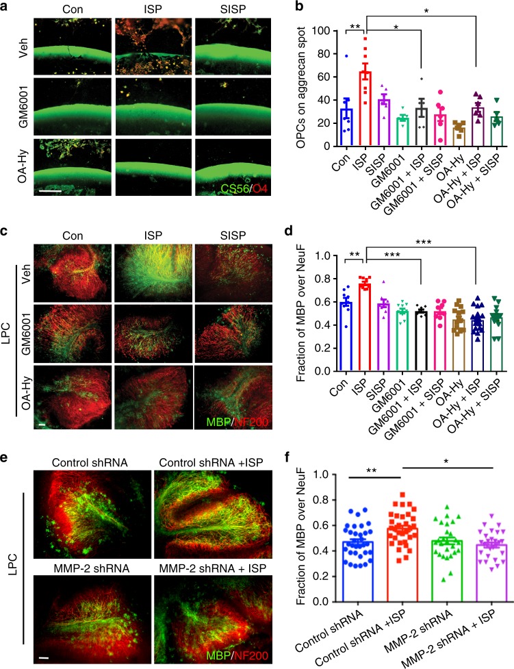
ISP-induced MMP-2 activity increases OPC migration and remyelination through CSPG degradation. a To ask whether ISP-induced protease activity is involved in OPC migration and remyelination, cultured OPCs were plated onto coverslips with CSPG spot gradients and treated with vehicle control, 2.5 μM ISP or SISP, 25 μM GM6001±ISP or SISP, or 100 nM MMP-2 inhibitor (OA-Hy)±ISP or SISP. Scare bar = 100 μm. b The amount of O4+ OPCs crossing the CSPG barrier was counted and quantified. ISP treatment (N = 37 spots, 5 replicates) significantly induced greater O4+ OPC migration past the CSPG barrier compared to control (One-Way ANOVA, Tukey’s posthoc test, P = 0.0002, F(11,48) = 5.013), GM6001 + ISP (N = 20 spots), or OA-Hy + ISP (N = 20 spots) treatments. N(Spots) = 31 Control, 26 SISP, 18 GM6001, 20 GM6001 + SISP, 18 OA-Hy, 19 OA-Hy + SISP). c To test whether remyelination was affected by protease inhibition, P7-9 cerebellar slices were all treated with LPC for 18 h, then treated with control vehicle, 2.5 μM ISP or SISP, 25 μM GM6001± ISP, or 100 nM OA-Hy±ISP for 9 days before staining for neurofilament (NF200) or MBP. Scale bar = 100 μm. d Remyelination was quantified through MBP and neurofilament colocalization. ISP treatment (N = 35 images from 4 replicates with up to 13 sections total) significantly increased MBP-neurofilament colocalization over control (One-Way ANOVA, Tukey’s posthoc test, P = 0.0001, F (8, 88) = 13.61), GM6001 + ISP (N = 28), and OA-Hy + ISP (N = 23) groups. N(images) = 17 Control, 22 SISP, 48 GM6001, 20 GM6001 + SISP, 22 OA-Hy, 22 OA-Hy + SISP. e, f Cerebellar slice cultures were treated with lentiviral constructs for 48 h before LPC treatment. Vehicle or ISP (2.5 μM) treatment followed for 6 days in vitro. Scale bar = 100 μm. MBP immunofluorescence (green) was then quantified with NF200 (red) (One-Way ANOVA, Tukey’s posthoc test. P = 0.0005. F(3, 116) = 6.395. N = around 30 images from 10 slices). The data are presented as mean ± s.e.m. *P < 0.05, **P < 0.01, ***p < 0.001

