Figure 3.
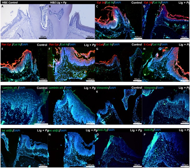
P. gingivalis-induced experimental periodontitis. (A) Histological hematoxylin-eosin staining of periodontium of non-treated site on the first molar of mice (Hematoxylin & Eosin) (10x magnification). (B) Morphological characteristics of periodontium of P. gingivalis ligature treated site on the first molar of mice (Hematoxylin & Eosin) (10x magnification). (C–H) Immunofluorescence staining for localization of Cytokeratin 14, Pan Cytokeratin and E-cadherin expression in epithelial tissue (red) and Collagen IV expression (green) in the periodontium (10x magnification). (I–P) Immunofluorescence staining for localization of Integrin β1, vimentin, laminin α5 and anti-P. gingivalis expression in the periodontium (green) at diseased site (10x magnification: I–M and O; 20x magnification: N,P). In all immunofluorescence images, nucleus has been stained by DAPI in blue.

