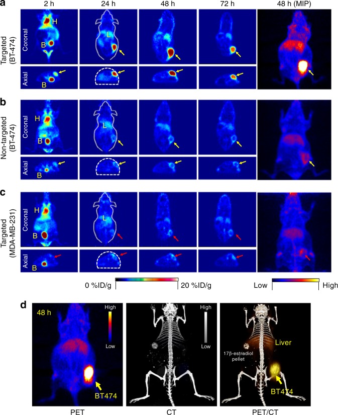
Fig. 3.
In vivo HER2-targeted PET imaging in xenograft breast cancer models. Serial coronal and axial tomographic PET images acquired at 2, 24, 48, and 72 h post i.v. injection of radiolabeled particle immunoconjugates in groups of tumor-bearing mice (N = 5 for each group) as follows—a targeted group: 89Zr-DFO-scFv-PEG-Cy5-C’ dots in BT-474 mice, b non-targeted group: 89Zr-DFO-Ctr/scFv-PEG-Cy5-C’ dots in BT-474 mice, and c targeted group: 89Zr-DFO-scFv-PEG-Cy5-C’ dots in MDA-MB-231 mice. For each group, maximum intensity projection (MIP) images were also acquired at 48 h p.i. H: heart, B: bladder, L: liver. d Representative MIP PET, CT, and PET/CT fusion images of 89Zr-DFO-scFv-PEG-Cy5-C’ dots in a BT-474 tumor-bearing mouse. All BT-474 tumors were marked with yellow arrows, while all MDA-MB-231 tumors were marked with red arrows

