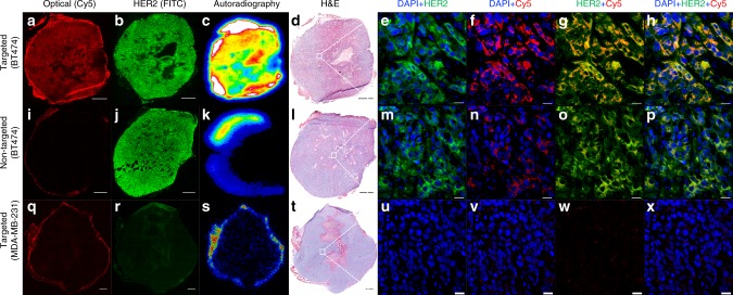
Fig. 5.
Correlative ex vivo tumor histopathology. From left to right: Cy5-fluorescence microscopy (scale bar: 1 mm), HER2 immunohistochemical staining (scale bar: 1 mm), autoradiography, H&E stained tumor tissue specimens (scale bar: 1 mm) harvested 72 h p.i., and confocal microscopy (scale bar: 20 μm) of random areas (white squares) from corresponding tumor tissue specimens. a–h Targeted group: 89Zr-DFO-scFv-PEG-Cy5-C’ dots, BT-474 tumor, i–p non-targeted group: 89Zr-DFO-Ctr/scFv-PEG-Cy5-C’ dots, BT-474 tumor, and q–x targeted group: 89Zr-DFO-scFv-PEG-Cy5-C’ dots, MDA-MB-231 tumor

