Fig. 6.
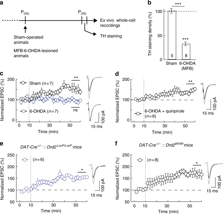
eCB-tLTP does not depend on D2R expressed by dopaminergic neurons. a–d Experiments of plasticity expression in 6-ODHA-ablated dopaminergic cells in the MFB. a Scheme of the protocol showing the time course of the in vivo 6-OHDA lesions and ex vivo electrophysiological recordings. b Unilateral 6-OHDA injection in MFB led to degeneration of dopaminergic nigral neurons, illustrated by a loss of their striatal afferences as illustrating by the summary bar graph of TH immunostaining quantification. Sham-operated rats display equivalent TH staining in both striata. c 10 post-pre pairings induced tLTP in sham-operated (n = 7, 7/7 cells showed tLTP) rats whereas tLTP was impaired in 6-OHDA-lesioned rats (n = 7, 1/7 cells showed tLTP), and d rescued with a D2R agonist, quinpirole (10 µM, n = 6, 5/6 cells showed tLTP). e, f STDP expression in selective cKO for D2R expressed by dopaminergic neurons. e In mice in which D2R was specifically knocked-out in dopaminergic neurons (DAT-Cre+/−::Drd2LoxP/LoxP mice) tLTP was induced with 15 post-pre pairings (n = 6, 6/6 cells showed tLTP). f tLTP was observed in mice serving as the Cre driver control (DAT-Cre+/−::Drd2Wt/Wt mice, n = 8; 6/8 cells showed tLTP). Representative traces are the average of 15 EPSCs during baseline (black traces) and 45 min after STDP protocol (grey traces). Vertical grey dashed line indicates the STDP protocol. Error bars represent sem. *p < 0.05; **p < 0.01; ***p < 0.001; ns: not significant by Wilcoxon Signed rank test (b) or one sample t-test (c–f)

