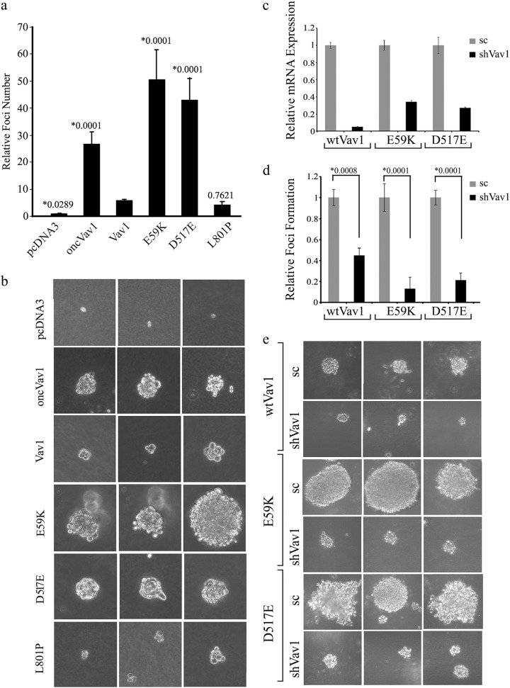
Fig. 5. Transforming activity of Vav1 mutants in vitro.
a NIH3T3 cells stably expressing pcDNA3, Vav1, oncVav1, E59K, D517E, and L801P were suspended in DMEM medium containing 0.3% agar and 10% calf serum and plated onto a bottom layer with 0.8% agar. 1 × 10⁵ cells were plated in a well in a 6-well plate in triplicate and the number of foci were counted 14 days later. The histogram presents means ± S.E. of triplicate values from three independent experiments. Unpaired Student’s t test (*) compare the number of foci obtained in each cell line to cells expressing Vav1. b Representative photographs of three foci from each transfected cell line in a are presented. c Vav1 was silenced in NIH3T3 cells stably expressing wtVav1, E59K, and D517E using shRNA sequences against wtVav1 (shVav1) or shscrambled (sc). The silencing efficiency of Vav1 was measured by real-time PCR using specific primers (supplementary Table 1). UBC and HPRT were used as a control. d NIH3T3 cells stably expressing Vav1, E59K, and D517E and cells in which Vav1 was silenced were analyzed for foci formation as detailed in a. The histogram presents means ± S.E. of triplicate values from two independent experiments. Unpaired Student’s t test was performed as indicated above. e Representative photographs of three foci from each cell line in d are presented

