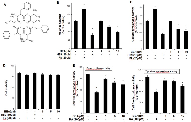
Figure 1.
The anti-melanogenic effect of beauvericin in B16F10 cells. (A) Chemical structure of beauvericin. (B) B16F10 cells were treated with beauvericin for 24 h. After harvesting, the cells were dissolved in a mixture of Soluene-350 and water. The melanin content was measured by absorbance at 500 nm (B). (C) B16F10 cells were treated with beauvericin for 24 h. After harvesting, the cells were lysed by sonication and assayed for cellular tyrosinase activity (dopa oxidase). Absorbance was immediately measured at 505 nm. Results were confirmed from at least three independent experiments, and values represent the means ± SEM. *P < 0.05 vs. untreated control. (D) Cell counting kit-8 was used to assay cell viability. Results were confirmed from at least three independent experiments, and values represent the means ± SEM. *P < 0.05 versus untreated control. (E) Cellular tyrosinase was isolated from B16F10 melanoma cells. After protein quantification, dopa oxidase activity and tyrosine hydroxylase activity activities were performed. The absorbance was measured spectrophotometrically at 505 nm and 475 nm, respectively. Data are presented as the means ± SEM of four independent experiments. Statistical significance of differences among the groups were assessed using the one-way analysis of variance (ANOVA) test followed by Tukey’s multiple-comparison test in the GraphPad Prism 5 Software. *P < 0.05 vs. control group. Fk, forskolin; KA: kojic acid; BEA, beauvericin.

