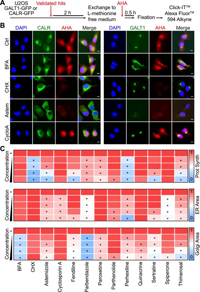
Figure 4.
Effects of confirmed secretion inhibitors on the ER and Golgi morphology and global protein synthesis. U2OS cells stably expressing CALR- GFP or GALT1-GFP were pre-treated with the selected secretion inhibitors (5, 10, 20, 40 μM) in L-methionine-free media then incubated with Click-iT® AHA for 30 minutes before subjected to a click reaction with Alexa Fluor® 594 alkyne (A). Images were acquired for quantification of nascent protein synthesis (cytoplasmic Alexa Fluor® 594 intensity), ER area (CALR-GFPhigh area), and Golgi area (GALT1- GFP bright area). BFA was used as positive control for Golgi disruption, and cycloheximide (CHX) was used as positive control for protein synthesis inhibition. Representative images of untreated controls, positive controls, as well as identified secretion inhibitors with typical ER disruption and/or protein synthesis inhibition (astemizole and cyclosporine A) are reported in (B), scale bar equals 10 μm. Quantitative data was normalized to untreated control and is summarized as heat map. Each block represents the mean value of 4 repeated measurements (C). Statistical analysis was performed by means of multiple t test, *p < 0.001 as compared to untreated controls.

