Figure 5.
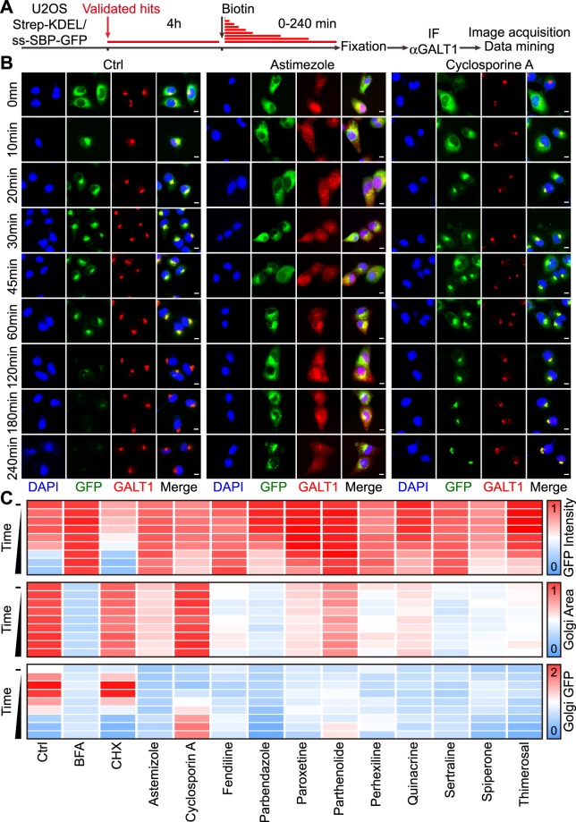
Effects of selected secretion inhibitors on conventional protein transport. U2OS cells coexpressing streptavidin-KDEL and ss-SBP-GFP were treated with selected agents at selected concentrations (20 μM for cyclosporin A, fendiline, parbendazole, paroxetine, parthenolide, quinacrine, sertraline, spiperone, thimerosal; 10 μM for astemizole and perhexiline) for 4 h, followed by biotin addition. Cells were fixed after different incubation periods, followed by immunofluorescence staining of GALT1 (A). Cytoplasmic GFP intensity was quantified as a means of protein secretion, GALT1bright area was quantified to indicate Golgi area and Golgi GFP intensity was used to measure the colocalization of GFP-tagged secretory cargo with the Golgi apparatus. Representative images of controls and astemizole or cyclosporine A treated cells are reported in (B); scale bar equals 10 μm. Quantitative data was normalized to untreated controls and summarized as heat map. Each block represents the mean value of 4 repeated measurements (C).

