Figure 3.
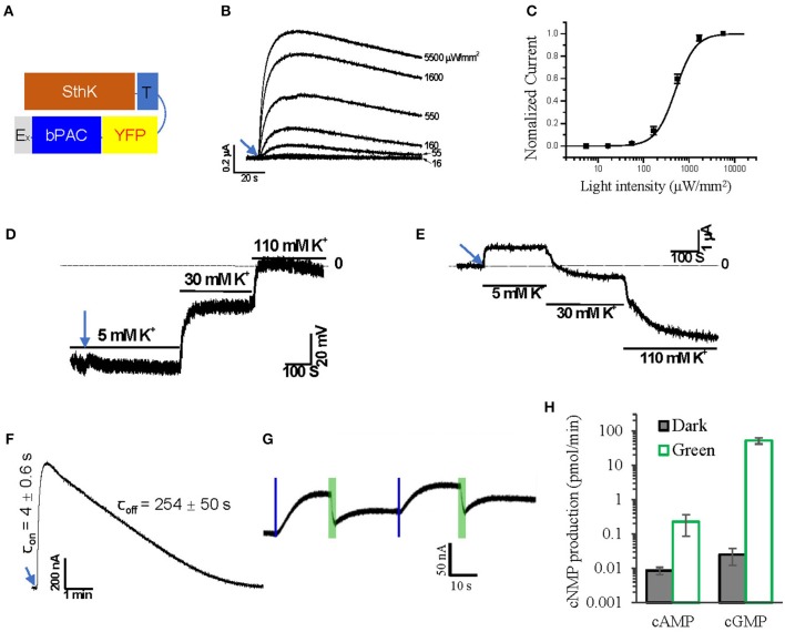
Design of a light-gated potassium channel. (A) Schematic of the designed SthK and bPAC fusion construct. (B) Representative Photocurrent traces recorded from Xenopus oocytes injected with 2 ng SthK-T-YFP-bPAC-Ex (SthK-bP) activated by 1 s blue light (473 nm) of different intensities, ~30 min recovery time in the dark for each illumination. (C) Normalized currents against light intensity curve fitted monoexponentially. The half-maximal light intensity value was determined to be 500 μW/mm2; n = 4, error bars = SEM. (D) Representative traces of membrane potential recording while switching bath solutions containing 5, 30, and 110 mM K+ after 5 s blue light (473 nm, 550 μW/mm2) illumination. (E) Representative traces of current recording (holding at −40 mV) while switching bath solutions containing 5, 30 and 110 mM K+ after 5 s blue light (473 nm, 550 μW/mm2) illumination. (F) On and off kinetics of SthK-bP photocurrents in oocytes with 1 s blue light (473 nm, 550 μW/mm2) illumination; for values, n = 4, error bars = SD. (G) Representative photocurrent traces from oocytes co-injected with 1.2 ng SthK-bP and 5 ng BeCyclop cRNA in Xenopus oocytes. Currents were induced by 200 ms blue light (473 nm, 550 μW/mm2) illumination and reduced by 3 s green light (532 nm, 1 mW/mm2) illumination. (H) cAMP and cGMP production of Xenopus oocyte membranes co-expressing SthK-bP and BeCyclop in the dark or light (532 nm, 80 μW/mm2). n = 3, error bars = SD.

