FIGURE 5.

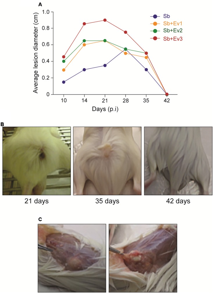
Progression of the subcutaneous infection in a murine model (A). Graph represents the evolution of the average diameter of skin lesions for 42 days resulting from subcutaneous fungal infection. A representative of 3 independent experiments, containing 5 animals each group at each timepoint. (B) Figures represent the external appearance of skin lesions formed at the fungus inoculation site, with the presence of ulcerative nodular formation at 21 days, nodular healing at 35 days, and complete regression at 42 days. (C) Internal appearance of a skin lesion at 35 days. A subcutaneous (left) encapsulated formation is observed, which when cut contains a purulent exudate (right).
