Figure 2.

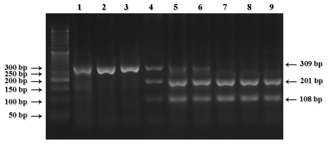
Pattern of 2% agarose gel electrophoresis of cytochrome P450 2D6*4 digested segments. Lanes 1–3 show homozygous samples for the mutant type (AA); lanes 7–9 wild-type samples (GG); and lanes 4–6 heterozygous samples (GA), respectively.

Pattern of 2% agarose gel electrophoresis of cytochrome P450 2D6*4 digested segments. Lanes 1–3 show homozygous samples for the mutant type (AA); lanes 7–9 wild-type samples (GG); and lanes 4–6 heterozygous samples (GA), respectively.