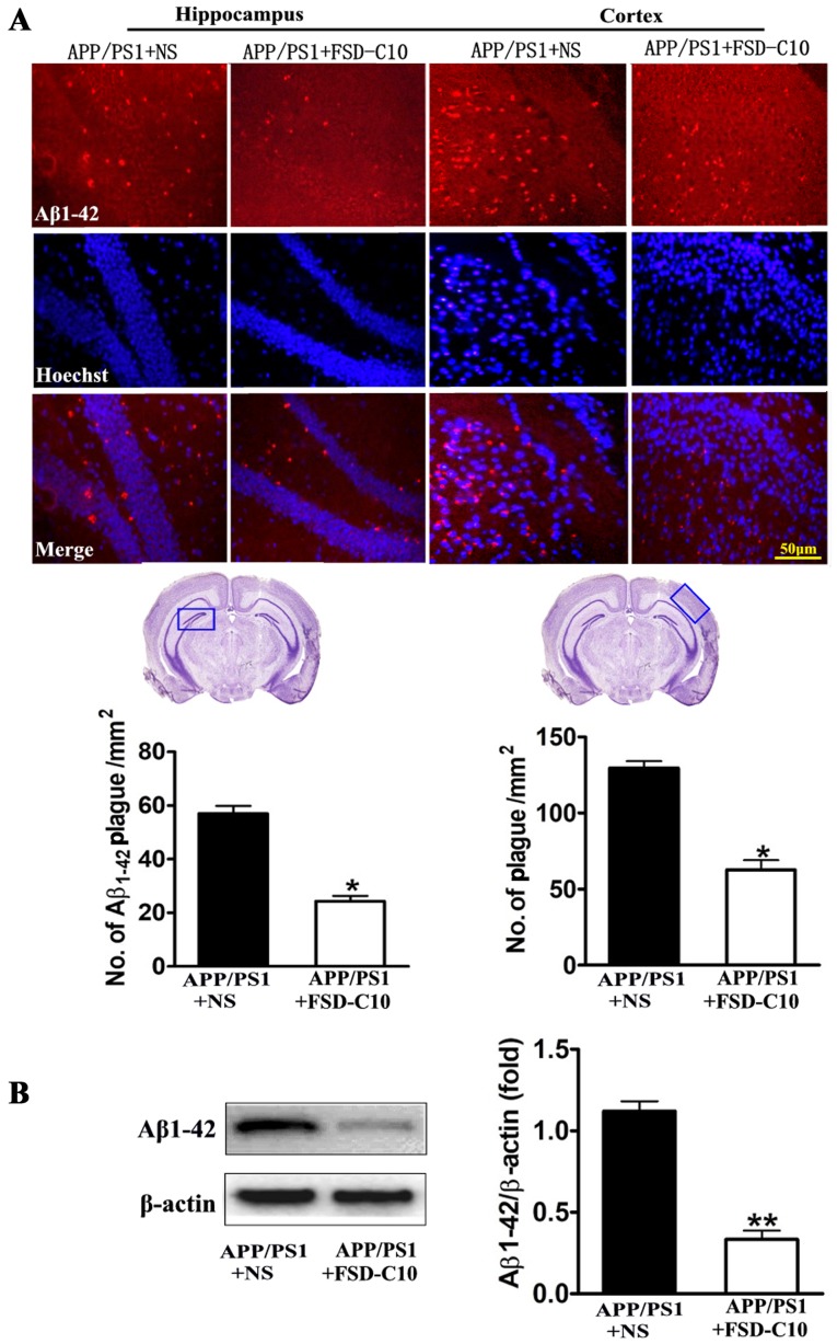
Figure 3.
FSD-C10 attenuates Aβ1–42 burden in hippocampus and cortex in APP/PS1 Tg mice. Aβ1–42 was measured by immunohistochemistry and western blot on two months after FSD-C10 administration. (A) Aβ1–42 expression was measured in hippocampus and cortex by immunohistochemistry and (B) in brain tissue by western blot. Quantitative results are shown as mean ± SEM of 4 mice each group, and one representative of three independent experiments. *P<0.05 and **P<0.01 vs. APP/PS1+NS.

