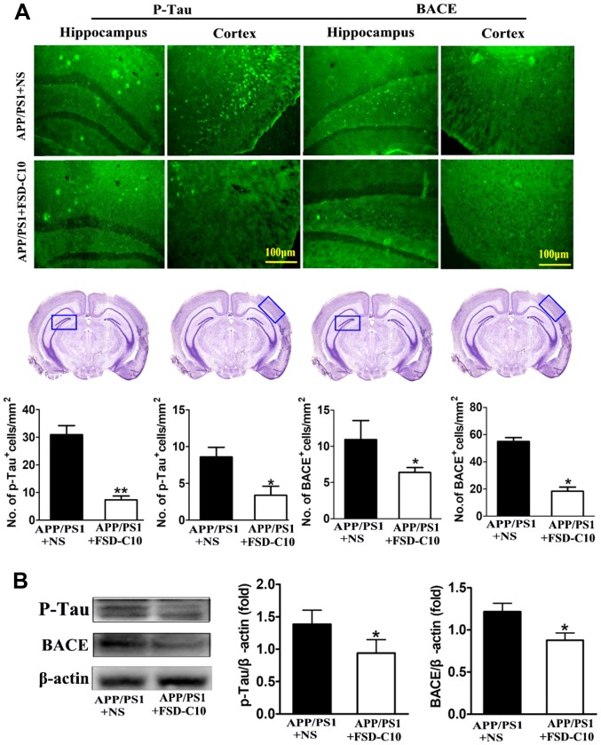
Figure 4.
FSD-C10 reduces Tau phosphorylation and BACE expression in hippocampus and cortex in APP/PS1 Tg mice. p-Tau and BACE were measured by immunohistochemistry and western blot two months after FSD-C10 administration. (A) The numbers of p-Tau and BACE positive cells in hippocampus and cortex were measured by immunohistochemistry and (B) their protein expression was measured in brain by western blot. Quantitative results are shown as mean ± SEM of 4 mice each group, and one representative of three independent experiments. *P<0.05 and **P<0.01 vs. APP/PS1+NS.

