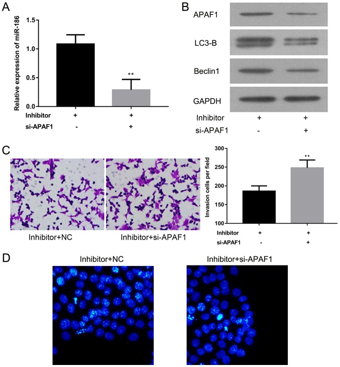
Figure 5.
Effect of APAF1 knockdown on the proliferation and apoptosis of A-431 cells. (A) The expression of miR-186 was examined by reverse transcription-quantitative polymerase chain reaction in A-431 cells with or without si-APAF1 in the presence of miR-186 inhibitors. (B) Western blotting was performed to detect the expression of APAF1, LC3B and Beclin1 proteins in A-431 cells treated with miR-186 inhibitors in the presence or absence of si-APAF1. (C) The invasive ability of A-431 cells treated with miR-186 inhibitors alone or in combination with si-APAF1 was measured using a Matrigel assay. Magnification, ×200. (D) Hoechst 33258 staining was performed to assess the apoptosis of A-431 cells transfected with miR-186 inhibitors with or without si-APAF1. Magnification, ×400. **P<0.01 vs. Inhibitor+NC. APAF1, apoptotic protease activating factor 1; LC3-B, light chain 3B; miR, microRNA; si, short interfering; NC, negative control.

