
Figure 3.
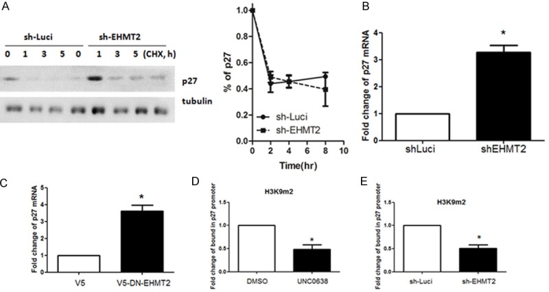
Knockdown of EHMT2 up-regulates p27 expression in a methyltransferase-dependent manner. A. Protein stability of p27 was detected in PANC-1 cell or PANC-1 sh-EHMT2 cells. Densitometry was utilized to quantify p27 protein levels after normalization with tubulin to obtain the percentage of p27 degradation (mean ± SD; n = 3). Error bars indicate SD. B. Expression of p27 in parental PANC-1 (sh-Luci) and EHMT2-deficient (sh-EHMT2) cells was determined by RT-qPCR analysis. Columns represent the mean of triplicate PCR assays, normalized to GAPDH (*P < 0.05). C. RT-qPCR analysis was used to investigate the expression of p27 in PANC-1 cells transfected with control or overexpressed methyltransferase-dead EHMT2 (DN-EHMT2). Columns represent the mean of triplicate PCR assays, normalized to GAPDH (*P < 0.05). D. PANC-1 cells were treated with DMSO or UNC0638. ChIP-qPCR analysis was performed to determine the status of H3K9m2 in the p27 gene promoter. Experiments were performed in triplicate. (*P < 0.05). E. ChIP-qPCR analysis was used to determine the status of H3K9m2 in the p27 gene promoter in PANC-1 or EHMT2 knockdown cells. The experiments were performed in triplicate (*P < 0.05).