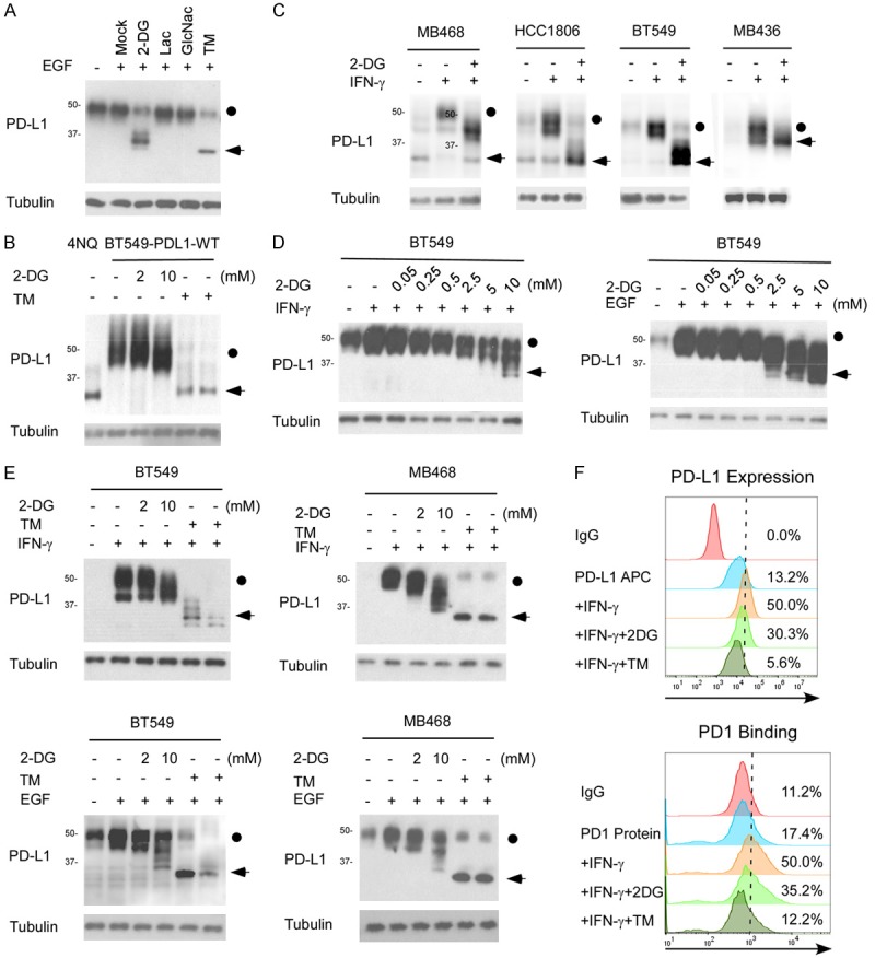
Figure 1.

2-DG deglycosylates PD-L1 in TNBC. A. Western blot analysis of PD-L1 protein expression in MDA-MB-231 cells treated with 2-DG, lactacystin (Lac), N-acetylglucosamine (GlcNac), or tunicamycin (TM). The dot indicates the glycosylated PD-L1 and the arrow indicates the deglycosyalted PD-L1. B. PDL1-overexpressing BT549 cells were treated with 2 or 10 mmol/L 2DG or with 1 µgml-1 TM for 12 hours and then subjected to immunoblotting with antibodies against PD-L1. BT549-4NQ cells were included as a negative control. C. Western blot analysis of PD-L1 protein expression in MB468, HCC1806, and BT549 cells treated with IFN-γ with or without 2-DG. D. BT549 cells were treated with indicated concentrations of 2-DG and IFN-γ or EGF for 12 hours, and then PD-L1 protein expression was determined by Western blot analysis. E. Western blot analysis of PD-L1 protein expression in MB468 and BT549 cells treated with 2 or 10 mmol/L 2-DG in combination with IFN-γ or EGF. TM treatment was used as the positive deglycosylation control. F. PD-L1 expression and PD-1 binding on the surface of BT549 cells were analyzed with FACS. The cells were treated with 10 mmol/L 2-DG, 1 µgml-1 TM, or 1 µgml-1 IFN-γ for 12 hours.
